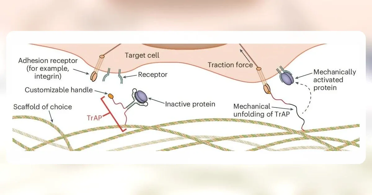
A new wound dressing is designed to wait for a repair cell before releasing biological signals. The experimental material collects healing proteins available locally or in blood preparations, keeps them inactive and responds when a cell pulls on the scaffold.
On July 27, 2026, researchers led by Imperial College London reported the system in Nature Materials after tests spanning injured rat femurs, surgically created skin defects in mice and human skin samples maintained in the laboratory. The active dressing reduced wound diameter in mice and improved integration with human tissue. No patients were treated in the study.
Cell Traction Unlocks Growth Factors at the Wound
Growth factors help coordinate cell movement, blood-vessel formation and tissue rebuilding. Delivering them as a treatment is difficult because proteins can break down quickly, while a large dose placed across a wound does not reproduce the timing or location of the body's own signals. The Imperial team approached that delivery problem as a mechanical one.
The material uses aptamer constructs called traction-force-activated payloads, or TrAPs, fixed to a collagen scaffold. Each construct binds a selected growth factor and keeps it from signaling. A repair cell adheres to the material and exerts traction as it moves. That force unfolds the construct, freeing the protein beside the cell rather than allowing it to diffuse from the dressing continuously.
The researchers loaded some scaffolds with growth factors and also tested empty constructs that collected molecules from injury sites or blood preparations. Across the experiments, the platform handled several proteins, including FGF-2, VEGF-A, PDGF-BB and HGF. The paper reports activity at doses orders of magnitude below current clinical standards, although lower experimental dosing does not by itself establish a safe or effective dose for patients.
The controls were central to the mechanism claim. Some sponges carried scrambled aptamers that had the same general material structure but could not perform the intended capture-and-release function. Others received a basic cysteine treatment. Differences between those controls and the active TrAP scaffold therefore tested whether force-responsive delivery added an effect beyond the collagen sponge alone.
Mouse Wounds and Donated Skin Answer Different Questions
For the skin pilot, the team created two 6 millimeter full-thickness wounds on each 12-week-old mouse and implanted collagen sponges immediately. Day-ten histology covered 32 wounds: nine with the basic control, 11 with scrambled aptamers and 12 with active TrAPs. The active group had a significantly smaller normalized wound diameter than either control group, while the two controls did not differ significantly from one another.
Those counts refer to wounds, not individual patients or a large clinical sample. Each mouse carried two wounds, and the researchers treated individual wounds as the unit of analysis. Researchers kept the mouse phase small under the approved animal protocol because it was an early cross-species check. Wounds were excluded when a sponge fell off during the study. That exclusion was disclosed, and it matters when interpreting a small experiment whose endpoint depends on the implanted material remaining in place.
The human evidence came from abdominal skin donated by three women aged 25 to 50 after abdominoplasty. Researchers made 3 millimeter wounds in the samples and maintained the tissue outside the body for four or eight days. The main experiment assessed four or five wounds from each donor in every treatment group. Those repeated samples improve within-donor comparisons, but they do not turn three donors into a large human population. Active multi-protein constructs increased measured overlap between cells and the sponge and improved dermis-sponge integration.
Ex vivo human tissue can show biological compatibility, but it cannot reproduce a patient's circulation, immune response or daily wound care. A separate rat femur model added evidence that the constructs remained functional in another injury environment and promoted vascularization. Taken together, the models support the proposed release mechanism across several tissues. They do not establish that the dressing closes chronic ulcers, burns or traumatic wounds in people.
Clinical Proof Is the Dressing's Hardest Test
The study's strongest result is not a finished treatment. It is the controlled comparison showing that a scaffold can keep captured signals inactive until cells apply force, then produce measurable effects in living animal tissue and donated human skin. The scrambled-aptamer groups make that explanation more persuasive than an uncontrolled observation of a wound becoming smaller.
The translational gap is still substantial. A clinical program would have to establish manufacturing consistency, safe protein capture and release, reliable attachment in real wounds and a benefit over current care. It would also need to test patients whose wounds may involve infection, impaired circulation or diabetes, conditions that were not reproduced by the reported mouse pilot or the donated-skin model. Imperial is developing the work through a proposed spinout, Traxion Biotech, but commercial activity is not clinical evidence.
Cell-triggered delivery may eventually solve a genuine timing problem in wound medicine: how to put a repair signal where it is needed without flooding the whole site. This paper shows that the mechanism can operate in demanding biological environments. Until patient trials show safer or better healing, however, the advance belongs to materials science, not to the clinic.